(02) 9484 7418
180 Pennant Hills Road
,
Thornleigh
,
NSW 2120
Book Online
About
Services
Pet Care
Desexing your Pet
General Vet Care
Health checks
Nurse Consults
Pets & Worms
Senior Pets
Pet Health
In House Blood Tests
Orthopaedic Surgery
Pet Cancer Care
Pet Dental Care
Scans & Imaging
Soft Tissue Surgery
Specialist Vet Referrals
Ultrasounds
X-ray and Radiology
Pet Wellbeing
Behaviour Consultations
Cat Boarding & Day Care
Dog Boarding
Doggy Daycare
Effective Flea Control
Microchipping
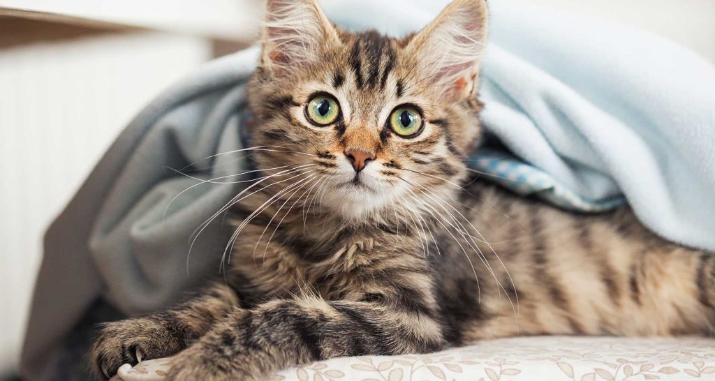
New Kitten
New Puppy
Pet Vaccination
Prescription Food & Diet
Emergency
Best for Pet
Pet Advice
Contact
Book Online
(02) 9484 7418
Book Online
Category:
Pet Advice
Search for:
Looks good!
Please enter a search query.
Read More
Pet Advice
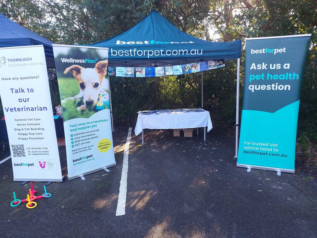
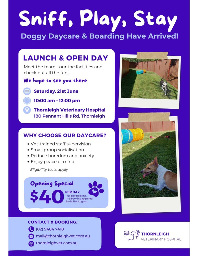
Doggy Daycare Grand Opening
Read More
Pet Advice
Doggy Daycare Opening
Read More
Pet Advice
What is desexing?
Read More
Pet Advice
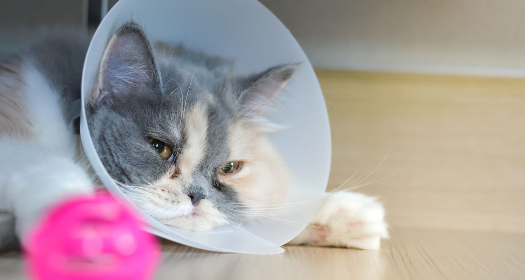
Cat bite abscesses
Read More
Pet Advice
Anal Gland Issues
Read More
Pet Advice
Eosinophilic granulomatous complex
Read More
Pet Advice
Ringworm
Read More
Pet Advice
Osteoarthritis in dogs
Read More
Pet Advice
Caring for your New Puppy
1
2
...
15